Ectopic Pregnancy
Ultrasound imaging is a varied field. In addition to its most widely known usage in pregnant women, it can detect a number of serious conditions in the human body. When compared and contrasted with MRI, X-Ray and CT scans, ultrasound is a an extremely useful imaging modality with its own helpful and unique diagnostic techniques. There are many common life-threatening conditions in the US population it can detect, including:
1. Cancer
Ultrasound imaging can show pictures of some soft tissue diseases that don’t show up clearly on x-rays. It's also an excellent imaging modality to distinguish fluid-filled cysts from solid tumors. It can be done quickly, and has an advantage over other imaging techniques because it doesn't expose patients to radiation.
Doctors often use ultrasound to do a needle-guided biopsy where by watching the screen, they can see the needle moving into the tumor. In addition, doppler flow machines can highlight the blood flowing through vessels. This is helpful because normal tissue blood flow images differently than blood flowing through a tumor. Color Doppler makes is easy for doctors to discover if cancer has spread into blood vessels, most notably in the liver and pancreas.
2. Heart Disease
During an echocardiogram, a transducer sends waves to bounce off of the heart. The resulting picture can detect heart disease, thus making ultrasound imaging an excellent tool for this type of life-threatening condition. Stress echocardiograms are performed while the heart is under physical stress due to exercising. Determining the heart's stress level while exercising can be key to getting a patient's heart disease under control.
3. Stroke
Carotid ultrasound is performed to evaluate a patient's risk of a stroke. The blood is monitored through the carotid arteries in the neck to check for stenosis. If ultrasound imaging detects stenosis, a stroke could be imminent.
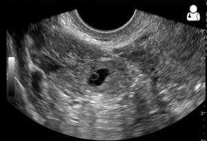
4. Ectopic Pregnancy
Ectopic pregnancy results when a fertilized egg implants outside the uterus. It is the number one cause of first-trimester maternal death. An ectopic pregnancy that isn't diagnosed and treated immediately could result in a ruptured fallopian tube, causing severe abdominal pain and bleeding. Permanent tube damage or even death could result, which is why an ultrasound 8-10 weeks into pregnancy is critical.
5. Enlarged Heart
Finally, ultrasound imaging can be used to diagnose an enlarged heart. An enlarged heart, if it goes undetected, can cause heart failure, blood clots, or cardiac arrest. It's important to get an ultrasound if shortness of breath, abnormal heart rhythm, and swelling in the body occur at the same time.
As you can see, ultrasound imaging is a critical tool in the diagnostic world. It has an invaluable place in hospitals and clinics all over the US.
About the Author
Brian Gill is Probo Medical’s Vice President of Marketing. He has been in the ultrasound industry since 1999. From sales to service to customer support, he has done everything from circuit board repair and on-site service to networking and PACS, to training clinicians on ultrasound equipment. Through the years, Brian has trained more than 500 clinicians on over 100 different ultrasound machines. Currently, Brian is known as the industry expert in evaluating ultrasounds and training users on all makes and models of ultrasound equipment, this includes consulting with manufacturers with equipment evaluations during all stages of product development.


